
06 Sep Endoscopic Diagnosis of Esophageal Varices
Endoscopic Diagnosis of Esophageal Varices
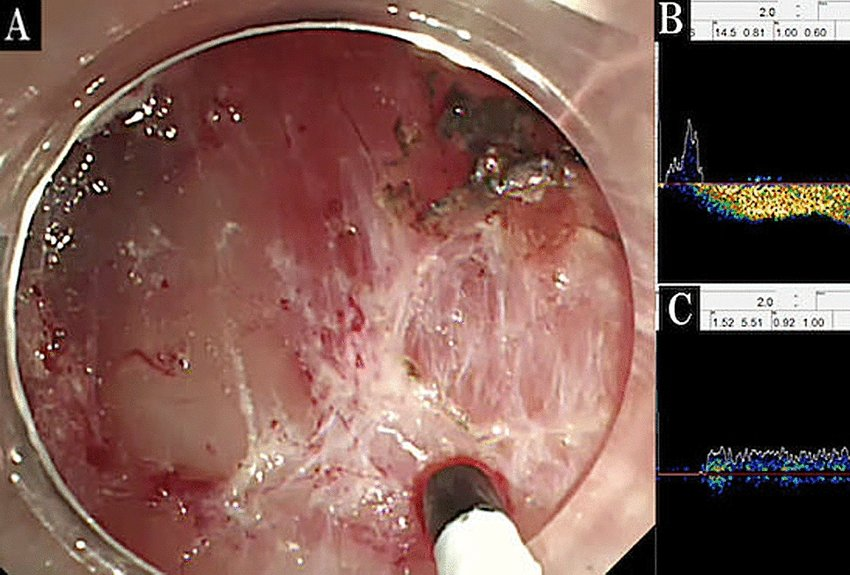
Examination with 16 MHz Doppler probe, passed through the endoscope, accesses the hemodynamic status of varices and evaluates the direction, waveform and amount of blood flow . Endoscopic diagnosis with 16 MHz is safe and allows identification of higher-risk varices, detection of penetrating varices, support of appropriate hemostasis, and evaluation after hemostatic treatment.
We are thrilled to speak with Dr. Yasutoshi Shiratori, Sherbrooke University Hospital, Quebec, Canada, about his impact studies using TCD Doppler systems in endoscopic procedures.
Dr. Shiratori is working as Advanced Therapeutic Endoscopy Fellow at Sherbrooke University Hospital in Quebec, Canada.

Dr. Shiratori, in your publications you deal with endoscopic diagnosis of esophageal varices and their evaluation. What particularly motivates you in your research?
Dr. Shiratori: “Esophageal variceal bleeding is a life-threatening complication of cirrhosis. Endoscopic variceal ligation is performed for urgent hemostasis and prophylactic treatment of esophageal varices. However, the probability of recurrence of variceal bleeding within one year is about 60% if patients do not receive appropriate treatment.”
You use Doppler ultrasound with the 16 MHz probe in your diagnostics. Why do you use this particular method?
Dr. Shiratori: “Until now, Doppler assessment was performed with endoscopic ultrasound to assess the hemodynamic status of varices. However, Doppler ultrasound evaluation is a complex technique with convex transducers that is unsuitable for routine clinical use and necessitates reinsertion of the endoscope when additional endoscopic variceal ligation is required.”
You say in your publications that the Doppler method is superior to the ultrasound method. What leads you to make this statement?
Dr. Shiratori: “Since the Doppler method is simple, it provides a straightforward view compared to the ultrasound method. In addition, Doppler ultrasound is simple, non-invasive and does not require replacement of the endoscope for additional treatment. Another advantage is that vascularity can be confirmed immediately afterwards. Older Doppler systems were indeed very limited, being audio-only Doppler devices. Therefore, an objective assessment of vascularity, such as direction and velocity of blood flow, could not be determined.
In the latest method, Doppler probe ultrasound examination with DWL’s systems, a Doppler probe passed through the endoscope evaluates the direction, waveform (to distinguish between arteries and veins), and amount of blood flow.”
If I understand you correctly, do you think that the method you presented can make a valuable contribution to safety, both for the method itself and for the patient being examined, when used with the endoscope?
Dr. Shiratori: “In our studies, we hypothesized that variceal velocity is associated with high-risk endoscopic varices due to portal hypertension, and therefore, the assessment of variceal velocity can be used to identify risks in the objective evaluations of varices, which then supports the improvement of the procedure. Therefore, the aim of our studies was to evaluate variceal velocities using the Doppler ultrasound method and to describe its feasibility.”
Recently you published a comparative study “Evaluation and management of esophageal varices by through-the-scope endoscopic Doppler probe method” in which you performed your new Doppler method called DOP on your patients. Would you say that the DOP method can have a positive impact on patient safety?
Dr. Shiratori: “The procedure in the studies is safe and includes identification of higher-risk varices, detection of penetrating varices, support of appropriate hemostasis, and evaluation after hemostatic treatment. However, we will conduct prospective studies to validate our findings.”
Dr. Shiratori, thank you very much for taking your time and wish you all the best for your future. We are looking forward to hearing about your further research.
Literature:
*Esophageal variceal treatment using a novel Doppler probe method; 2021
Shiratori, Y., Yoshimoto, T., & Yamamoto, K;:
**Evaluation and management of esophageal varices by through-the-scope endoscopic Doppler probe method; March 2022
Yasutoshi Shiratori, Kazuki Yamamoto, Shuhei Okuyama, Takaaki Yoshimoto, Takashi Ikeya, Syuichi Okada,Katsuyuki Fukuda, George Rateb
***Doppler probe method to reduce delayed bleeding after endoscopic submucosal dissection in the stomach: a propensity-score matched study (with video); May 2022
Yasutoshi Shiratori, Takashi Ikeya,. Kazuki Yamamoto, Ayaka Takasu, Yuichirou Suzuki, Syuichi Okada, Katsuyuki Fukuda, George Rateb;Ma