
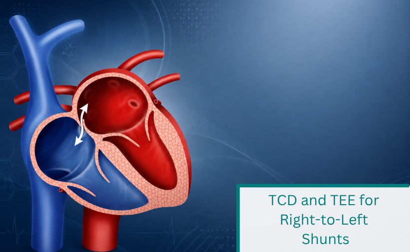
03 Aug TCD and TEE for Right-to-Left Shunts
Almost Perfect Concordance Between TCD and TEE in Right-to-Left Shunt Quantification
Contrast-enhanced transcranial Doppler (TCD) offers a non-invasive approach for the detection of right-to-left shunts associated with patent foramen ovale (PFO). In this study, Belvís et al. investigated how closely TCD agrees with transesophageal echocardiography (TEE), the established reference method, when both examinations are performed simultaneously.
Clinical background
A patent foramen ovale is present in approximately 30% of the adult population and occurs more frequently in patients with cryptogenic cerebral infarction. Several hypotheses have been proposed to explain the association between PFO and stroke, including paradoxical embolism, arrhythmogenic mechanisms, and in situ thrombus formation.
TEE and TCD are considered the methods of choice for PFO detection. Discrepancies between both techniques have previously been attributed to differences in hemodynamic conditions, the performance of the Valsalva maneuver, and the fact that examinations were not conducted simultaneously. Prior to this study, no systematic analysis of RLS quantification concordance between TCD and TEE had been performed.
Study Objective
The objectives of the study were to:
- Perform TCD and TEE simultaneously
- Compare PFO detection rates between both methods
- Analyze the concordance of RLS quantification between TCD and TEE
Methods
A total of 110 consecutive stroke patients undergoing TEE for etiological evaluation were prospectively included. At the same time, a contrast-enhanced TCD study was performed following the protocol of the Venice Consensus Conference.
Examination Protocol
- Contrast agent: agitated mixture of 9 mL isotonic saline and 1 mL air
- Injection site: right antecubital vein
- Valsalva maneuver: initiated 5 seconds after contrast injection and maintained for 10 seconds
- Two contrast injections were performed
- RLS quantification was based on the second injection
A PFO was diagnosed when microbubbles were detected in the left atrium within three cardiac cycles after right atrial opacification on TEE, together with corresponding microbubble signals in the middle cerebral artery on TCD.
RLS Quantification Criteria
TEE
- Small: 3–10 microbubbles
- Moderate: 11–30 microbubbles
- Large: > 30 microbubbles
TCD
- Minimum: 1–10 microbubbles
- Moderate: 11–25 microbubbles
- Massive: > 25 microbubbles, including shower or curtain patterns
Results
- Mean age: 56.7 years
- 60.9% male
- 70.9% cryptogenic Strokes
PFO Detection
- After the first contrast injection:
- TCD detected PFO in 30% of patients
- TEE detected PFO in 31.8% of patients
- After the second injection:
- Both methods identified the same patients (32.7%)
Missed detections were limited to patients with small shunts.
RLS Qualification
The concordance between TCD and TEE was almost perfect:
- Cohen’s Kappa: 0.928 (p = 0.001)
- Only 4 discrepancies among 110 patients
In patients with cryptogenic stroke, concordance remained almost perfect (Kappa = 0.93).
Conclusion
The high concordance between transcranial Doppler (TCD) and transesophageal echocardiography (TEE) (Cohen’s Kappa ≈ 0.93) suggests that discrepancies reported in earlier studies are likely related to non-simultaneous testing and variability in hemodynamic conditions or Valsalva maneuver execution. Differences between methods were mainly observed in cases of small shunts, while moderate and large shunts showed consistent classification across both modalities. Overall, the study confirmed an almost perfect concordance between the non-invasive TCD and the invasive TEE for the detection and quantification of right-to-left shunts.
Belvís, R., Leta, R. G., Martí-Fàbregas, J., Cocho, D., Carreras, F., Pons-Lladó, G., & Martí-Vilalta, J. L. (2006). Almost perfect concordance between simultaneous transcranial Doppler and transesophageal echocardiography in the quantification of right-to-left shunts. Journal of Neuroimaging, 16(2), 133–138. https://doi.org/10.1111/j.1552-6569.2006.00021.x